I am a Senior Lecturer in human anatomy at the University of Liverpool with research projects in the area of functional morphology and biomechanics. If you are interested in a PhD in this field, please contact me so we can discuss funding. Some potential funding options can be found on the UoL Website here.
Some of my current and previous work is highlighted below as well as on ResearchGate.
Some of my current and previous work is highlighted below as well as on ResearchGate.
Temporal fascia function during human growth: biomechanical modelling to predict the impact of surgical intervention
This BBSRC funded project (2023-2026) aims to explore the function of the temporal fascia, characterising its role during craniofacial growth and mastication. There is growing evidence of the mechanical significance of the temporal fascia during chewing, in controlling temporalis muscle force direction and bone strain distribution over the zygomatic arch and cranial vault. However, it is often overlooked in investigations of jaw muscle and temporomandibular joint (TMJ) function during mastication, resulting in a lack of knowledge about its role in regulating cranial strain and joint reaction forces. In addition, there is evidence that the temporal fascia impacts cranial ontogenetic development and zygomatic arch shape; however, the consequence of detaching the fascia during surgery is unknown for normal skull function, and healthy bone growth in children or adults.
Evolution of unique cranial morphology and function in lagomorphs
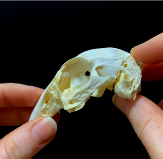
This is a funded NERC Doctoral Training Program of which I am the primary supervisor for Amber Wood-Bailey. Rabbits and hares (leporids, lagomorphs) are unique among mammals for having a joint within the cranium that provides movement, rather than having a completely rigid skull. This movement, or cranial kinesis, is common in vertebrates such as reptiles, but has not evolved in any other mammals, so its evolution and function is not well understood. Early studies have suggested that cranial kinesis in rabbits functions as a shock-absorbing mechanism to dissipate kinetic energy during impacts associated with fast running. However, this has not been tested. In this project, Amber will take an evolutionary comparative approach to explore how the biomechanics, ecology, and environmental factors influence the evolution of lagomorph skull shape and function, through the application of sophisticated computational methods such as finite element analysis, biplanar x-ray videography and geometric morphometrics.
Wood-Bailey AP, Cox PG, and Sharp AC (2022) The evolution of unique cranial traits in leporid lagomorphs. PeerJ, 10:e14414. 10.7717/peerj.14414
Cranial sutures and craniosynostosis
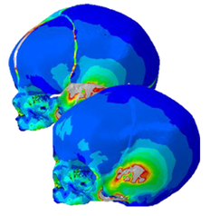
The primary aim of this research is to widen clinical knowledge on the influence of premature sutural obliteration on the shape of the calvarium and facial skeleton both pre- and post-operatively. Given that social anonymity remains a key measure of outcomes assessment, we will investigate the link between calvarial expansion and facial symmetry up to sixteen years of age. Furthermore, employing an increased duration for follow-up could identify markers for re-synostosis and the possibility of surgical recurrence, an area of paucity in current literature.
Secondarily, this project aims to advance knowledge of the biomechanical role of sutures during growth and the transitions between suckling to chewing. This project is being lead by PhD student Emily Baxter and is in collaboration with clinicians at Alder Hey Children's Hospital.
Hulls L, Moazen M and Sharp AC (2022) Finite element analysis of infant brain expansion and muscle activation shows the importance of cranial sutures for healthy skull growth. Anatomical Society Summer Meeting, Dublin, Ireland. Poster: Download here.
Baxter E, Moazen M, Sharp A (2024) Building a finite element model to simulate physiologic loading with metopic and unilateral coronal suture fusion in an infant skull. Anatomical Society Winter Meeting, Liverpool, UK. Poster: DOI: 10.13140/RG.2.2.30938.72644
The role of soft tissues in cranial biomechanics
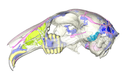
This BBSRC funded project (2015-2018) with Prof Susan Evans (UCL) and the University of Hull used a combination of traditional anatomical work and computational biomechanics (MDA and FEA) to understand the mechanical role and significance of non-muscular soft tissues in the reptilian and mammalian skull during feeding.
Figure: lateral view of a rabbit skull
Figure: lateral view of a rabbit skull
Sharp AC, Dutel H, Watson PJ, Gröning F, Crumpton N, Fagan MJ, and Evans SE (2023) Assessment of the mechanical role of cranial sutures in the mammalian skull: Computational biomechanical modelling of the rat skull. Journal of Morphology 284:e21555. DOI: 10.1002/jmor.21555
Watson PJ, Sharp AC, Choudhary T, Fagan MJ, Dutel H, Evans SE, and Gröning F (2021) Computational biomechanical modelling of the rabbit cranium during mastication. Scientific Reports, 11(1):13196. DOI: 10.1038/s41598-021-92558-5
Dutel H, Gröning F, Sharp AC, Watson PJ, Herrel A, Ross CF, Jones MEH, Evans SE, and Fagan MJ (2021) Comparative cranial biomechanics in two lizard species: impact of variation in cranial design. The Journal of Experimental Biology. DOI: 10.1242/jeb.234831
Jones ME, Gröning F, Aspden R, Dutel H, Sharp AC, Moazen M, Fagan MJ, and Evans SE (2020) The biomechanical role of the chondrocranium and the material properties of cartilage. Vertebrate Zoology 70:699-715. DOI: https://doi.org/10.26049/VZ70-4-2020-10
Human foot anatomy and function
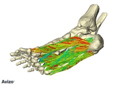
I also occasionally study the functional anatomy of the human foot. The human foot is unique among vertebrates and is characterized by a pronounced longitudinal arch (LA) that compresses and recoils in response to external load during locomotion. This has typically been considered a passive process; however, it has recently been shown that the plantar intrinsic foot muscles have the capacity to actively assist in supporting the LA. Through dissection and imaging I have investigated anatomical variation of the intrinsic foot muscles.
Funding: MRC DynPadAge Project - In vivo thickness and dynamic behaviour of the heel fat pad during ageing: biomechanical and anthropological impact of footwear with Dr Kris D'Aout (University of Liverpool), Dr Catherine Willems (University College Ghent), and Dr Claire Brockett (University of Sheffield).
Aland RC, and Sharp AC (2021) Anomalous plantar intrinsic foot muscle attaching to the medial longitudinal arch: possible mechanism for medial nerve entrapment: a case report. Journal of Medical Case Reports. DOI: 15:58. 10.1186/s13256-021-02676-x
Aland, RC, Koutsoukis, FJA, Bennett, M, Sharp, AC and Gosden, E (2017) Flexor digitorum brevis - segmentation, pennation and PCSA. Australian & New Zealand Association of Clinical Anatomists (ANZACA), Auckland, New Zealand.
3D Visualisation and Reconstruction
Using a combination of CT and MRI scanning, gross dissection and biomechanical modelling, we can visualize and investigate the function of a variety of soft- and hard-tissue structures.
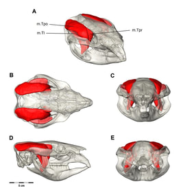
Figure: Digital dissection of the temporalis muscle group in the common wombat. 3D PDF available |
Information gathered from extant animals can then be compared to fossil specimens, allowing the reconstruction of muscles and other soft-tissue structures, which are rarely preserved in fossils.
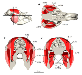
Figure: Digital reconstruction of the jaw adductor musculature in the Diprotodon |
Cranial Morphology, Biomechanics and Diet of Marsupial Herbivores
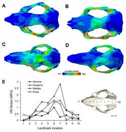
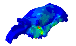
Marsupial herbivores come in many shapes and sizes, from hopping kangaroos to lumbering wombats. Studies on teeth, dentaries and jaw adductor muscles indicate that marsupial herbivores exhibit different specializations for grazing and browsing, including hypsodonty, molar progression and high bite forces. However, the relationships between skull morphology, biomechanical performance and diet are still relatively unknown. This research aims to investigate the interaction between biomechanical and non-biomechanical factors, including the lifestyle of the animal and its environment, in selection for skull morphology to meet multiple functional demands. In accordance with previous studies, results show the mammalian skull may not be optimized solely to resist forces generated during feeding.
Figure: Predicted distribution of stress across the cranial models of the (A) common wombat (V. ursinus), (B) koala (P. cinereus), (C) red kangaroo (M. rufus), and (D) swamp wallaby (W. bicolor).
Figure: Predicted distribution of stress across the cranial models of the (A) common wombat (V. ursinus), (B) koala (P. cinereus), (C) red kangaroo (M. rufus), and (D) swamp wallaby (W. bicolor).
Sinus Morphology of Marsupial Megafauna
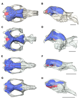
Cranial sinuses are air-filled cavities resulting from the resorption and deposition of bone through pneumatisation in response to biomechanical stress. The morphology of a pneumatic bone represents an optimisation between strength and being light weight. Marsupial megafauna are "airheads" in that most of their skull is composed of very large air sinuses, and their brains are relatively small compared to the overall size of the skull. The size and morphology of the sinuses changes with the size of the animal and the morphology of the skull. Using CT scans to reconstruct the skulls and estimate the volume of the sinuses and brains we can get a better understanding of their size, morphology and evolution.
Figure: Three-dimensional reconstructions of Diprotodon optatum (A-B), Zygomaturus tasmanicum (C-D), Neohelos stirtoni (E-F) and Propalorchestes sp. (G-H) showing the extent of the auditory, squamosal, parietal and frontal sinuses in blue, and brain endocast in red. Skulls are shown in dorsal (A, C, E, G) and lateral (B, D, F, H) views. Scale bars represent 10 cm.
Figure: Three-dimensional reconstructions of Diprotodon optatum (A-B), Zygomaturus tasmanicum (C-D), Neohelos stirtoni (E-F) and Propalorchestes sp. (G-H) showing the extent of the auditory, squamosal, parietal and frontal sinuses in blue, and brain endocast in red. Skulls are shown in dorsal (A, C, E, G) and lateral (B, D, F, H) views. Scale bars represent 10 cm.
Cranial Morphology and Function of Diprotodon
This project is work from my PhD. Diprotodon optatum is the largest marsupial known and became extinct about 45,000 years ago. Using CT scanning and an engineering technique called finite element analysis (FEA) I am investigating the structure and function of the extensive cranial sinuses. Another aspect of this project is predicting the bite force and feeding biomechanics to explore the diet and behaviour of these animals.
